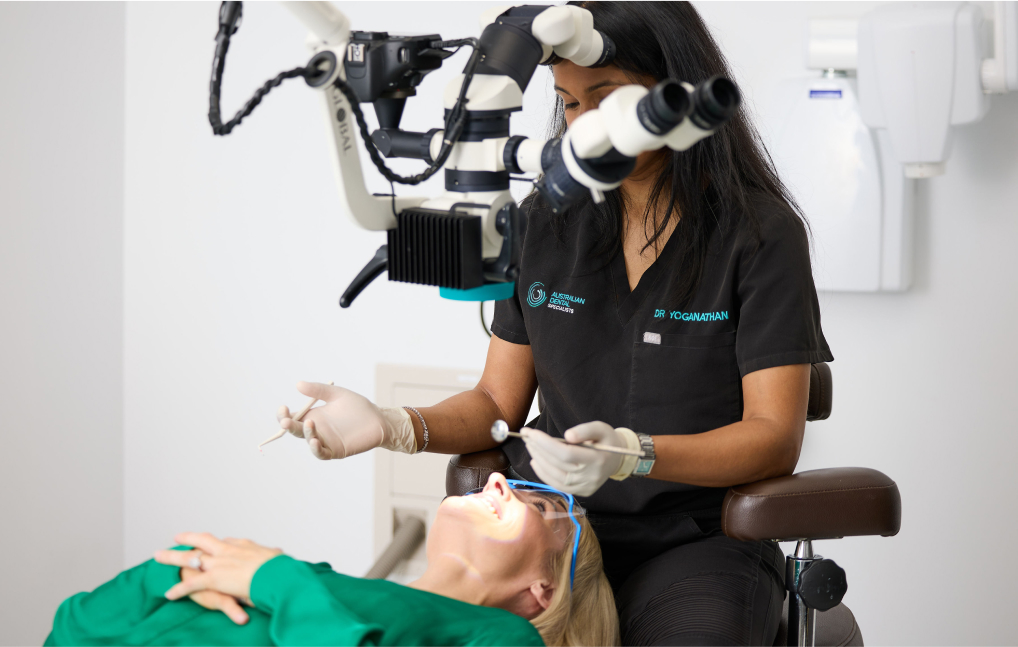
Microscopic root canal treatment uses up to 25 times magnification to uncover hidden canals and microscopic fractures, giving patients up to 2.9 to 3.2 times higher odds of a successful outcome compared with traditional methods. The dental operating microscope reveals what the naked eye cannot, which is why Dr Varayini Yoganathan and our specialist endodontic team use one for every procedure at Australian Dental Specialists.
The single most important factor in root canal success is whether every infected canal is found, cleaned, and sealed. Canals that are missed remain a reservoir for bacteria, and missed canals are the most common reason root canal treatment eventually fails. The smaller and more curved the canal, the harder it is to identify under standard lighting and ordinary loupes. Magnification at the level provided by a dental operating microscope brings these canals into view and gives the specialist a far better chance of treating the tooth completely the first time.
For patients, this matters because a missed canal usually shows up months or years later as persistent infection, a dull ache on biting, or a recurring abscess. By that point the tooth has often lost more structure, and retreatment, surgery, or extraction is the path forward. Magnification at the first appointment reduces that risk meaningfully.
This article explains what a dental operating microscope is, how 25 times magnification changes the procedure, what the published evidence shows about outcomes, and what this looks like in practice for patients treated at our Norwest clinic.
A dental operating microscope, or DOM, is a precision optical instrument purpose-built for dentistry. Unlike dental loupes, which typically magnify between 2.5 and 6 times, a DOM provides continuously adjustable magnification from 4 to 25 times, paired with integrated coaxial illumination.
That coaxial light is a critical distinction. The light source runs parallel to the clinician’s line of sight, shining directly into the canal without creating shadows. A headlamp clipped onto loupes casts shadows at depth and loses brightness as magnification increases.
| Magnification Range | Clinical Use |
| Low (2 to 8 times) | Locating canal openings, initial assessment, flap elevation |
| Mid (8 to 16 times) | Primary canal work, MB2 detection, detailed canal preparation |
| High (16 to 25 times) | Detecting microfractures, finding calcified canals, apical anatomy |
Your specialist moves between these ranges during a single procedure, zooming in for fine detail and out for broader orientation, all from an ergonomic seated position.
How widely microscopes are used
Among specialist endodontists, adoption is near universal. A 2023 global survey found that around 91% of endodontic association members use a dental operating microscope regularly, including members of the Australian and New Zealand Academy of Endodontists and the Australian Society of Endodontology. In Australia, most general dental practitioners have some magnification available, though most use loupes rather than microscopes. This difference in equipment partly explains why specialist endodontists achieve consistently higher success rates for complex cases.
Source: International Endodontic Journal, 2023. American Association of Endodontists, 2016
Finding hidden canals
The mesiobuccal 2 (MB2) canal in upper molar teeth illustrates why magnification matters. Present in up to 40% of upper molars, this canal becomes a reservoir for bacteria when left untreated, leading to persistent infection and treatment failure.
| Detection Method | MB2 Canal Detection Rate |
| Naked eye (no magnification) | 18 to 25% |
| Dental loupes (2.5 to 6 times) | 45 to 55% |
| Dental operating microscope (standard) | 57 to 70% |
| DOM with ultrasonic dentine removal | 85 to 89% |
Research using a staged detection approach found that direct vision alone identifies 36% of MB2 canals, the operating microscope alone identifies 54%, and the combination of microscope with selective dentine removal using ultrasonic tips raises detection to 72%. In specialist practice, where the complete protocol is applied, detection rates reach 85 to 89%. Every canal that goes undetected goes untreated, and untreated canals are the most common reason root canal treatment fails.
Source: PubMed Central, 2022
Detecting cracks and fractures
Cracked teeth present one of endodontics’ greatest diagnostic challenges. A hairline fracture can cause persistent pain or intermittent discomfort that mimics other conditions. Under normal examination, these cracks are invisible.
At high magnification with coaxial illumination, the microscope reveals structural defects that otherwise go undetected, including coronal cracks extending from the biting surface, radicular fractures along the root, and the distinction between harmless craze lines and true structural fractures. This precision prevents two costly errors: treating a tooth that cannot be saved, and extracting one that could have been preserved.
Source: Global Surgical Corporation, 2024
Navigating calcified and blocked canals
As teeth age, canals can become partially or completely blocked by mineral deposits. Under the microscope, your specialist identifies the subtle colour and texture changes marking a calcified canal’s location, including the characteristic small, dark brown dots showing where the canal has receded.
When combined with three-dimensional cone beam CT imaging and piezoelectric ultrasonic instruments, the microscope enables specialists to detect and navigate calcified canals that would otherwise be missed. CBCT provides pre-treatment localisation of the calcification, the microscope provides direct visualisation during treatment, and ultrasonic instruments allow precise dentine removal to expose the canal pathway. Research shows this combined approach successfully manages 85% or more of complex calcified canal cases that would otherwise often result in extraction.
Source: PubMed Central, 2016. PubMed, 2025
Non-surgical root canal treatment
A 2025 retrospective cohort study compared microscope-assisted and traditional root canal treatment in permanent posterior teeth.
| Measurement | Microscope-Assisted | Traditional |
| Odds of success (strict criteria) | 2.91 times higher | Baseline |
| Odds of success (loose criteria) | 3.25 times higher | Baseline |
| Pooled success rate (specialist practice) | 92 to 94% | 59 to 88% |
Patients receiving microscope-assisted treatment had nearly three times the odds of a successful outcome. This is particularly significant for posterior teeth, which have the most complex canal anatomy.
Source: PubMed, 2025. International Journal of Community Medicine and Public Health, 2023
Retreatment outcomes
When a previous root canal has failed, microscopic retreatment offers substantially better prospects than traditional techniques. Contemporary protocols using an operating microscope combined with nickel-titanium rotary files, ultrasonic irrigation, and thermoplastic obturation achieve success rates of 90 to 92% in recent cohort studies, a significant improvement over historical retreatment outcomes of 74 to 85%.
Source: Compendium of Continuing Education in Dentistry, 2025. Nature, 2024
Complex case success rates
The microscope’s advantage is most pronounced when complications arise.
| Complexity Type | Success Rate with Microscope |
| Broken instrument removal | 83 to 85% |
| Missed canal identification | 83% |
| Calcified canal management | 85% and above |
| Root perforation repair | 73 to 96% |
For broken instrument removal, visibility improvement alone increases success from 47.7% without magnification to 85.3% with the microscope.
Source: PubMed, 2025. PubMed Central, 2016
Surgical endodontics (apicoectomy)
The most dramatic difference appears in surgical cases.
| Surgical Approach | Success Rate |
| Endodontic microsurgery (with microscope) | 91 to 94% |
| Traditional apicoectomy (without microscope) | 59% |
A 35 percentage point improvement, representing a 60% relative risk reduction in treatment failure. Modern endodontic microsurgery under a microscope has become the standard of care for surgical retreatment, and is the approach used at Australian Dental Specialists for root end surgery.
Source: Nature, 2024. PubMed, 2005
Better tooth preservation
The microscope lets your specialist work with extraordinary precision, removing only what is necessary and preserving maximum healthy tooth structure. Less dentine removal during access, less exploratory work during canal negotiation. The result is a structurally stronger tooth, better equipped to support a crown and serve you for decades.
Fewer retreatments
With microscope-assisted treatment delivering 2.9 to 3.2 times the odds of success, you are far less likely to face the cost, time, and discomfort of having the procedure repeated. Specialist endodontic fees vary by tooth type, case complexity, and the experience of the treating clinician, and retreatment adds further cost and time. Getting it right the first time has genuine financial and practical value.
Source: Healthdirect Australia, Cost of Dental Care, 2024
Solving problems other methods cannot
Some clinical situations are only manageable with microscopic precision. Broken instruments lodged in canals see retrieval success improve from 47.7% to 85.3%. Calcified canals achieve 85% or higher success using the combined CBCT, microscope, and ultrasonic protocol. Root perforations show repair success of 73 to 96% under magnification. Failed previous root canals reach 90 to 92% success with contemporary microscopic retreatment.
If you have been told a tooth cannot be saved, a specialist assessment with microscope access may reveal options that were not visible during the initial examination.
Visual documentation
Modern dental microscopes integrate with digital imaging systems, capturing high-resolution photographs and video during treatment. You can see before-and-after images of your own treatment, your referring dentist receives detailed documentation for follow-up care, and a complete visual record exists for your file. This is particularly useful when your case involves multiple visits or when ongoing collaboration with your general dentist is part of the treatment plan.
Better diagnostic clarity at consultation
Magnification also changes the consultation conversation. Findings that would otherwise sit in clinical notes can be shown to you on a screen during your appointment. A hairline crack at the base of a tooth, a dark calcified canal entrance, or residual filling material from a previous treatment all become visible. That clarity helps you understand the recommendation and weigh it against alternatives such as extraction and implant replacement.
Less invasive access
The operating microscope allows the specialist to keep access cavities small. A smaller opening preserves more of the original tooth structure, which directly affects how strong the tooth will be after the crown is fitted. Conservative access, made possible by precise visualisation, is one of the quieter ways the microscope improves long-term outcomes.
Every root canal procedure at Australian Dental Specialists is performed under a Zeiss dental operating microscope as standard care, not as an add-on. Dr Varayini Yoganathan pairs microscope visualisation with 3D CBCT imaging and an assistant-side optic, so the treating team and the dental assistant share the same magnified view throughout the procedure, reducing the time spent repositioning or re-explaining findings mid-treatment.
Where clinically appropriate and with patient consent, microscope and CBCT findings are captured as part of the clinical record. This means your referring dentist receives clear documentation of exactly what was found and treated, and you can see the same images during your consultation rather than relying on a verbal description alone. For complex cases referred to us, including calcified canals, broken instruments, and previous failed treatment, this combination of microscope, CBCT, and specialist training is the basis for the case assessment you receive at your first visit.
What is microscopic root canal treatment?
Root canal therapy performed using a dental operating microscope that magnifies the treatment area up to 25 times. Combined with shadow-free coaxial illumination, it allows your endodontist to see anatomical details invisible to the naked eye, including hidden canals, microfractures, and calcification.
Does microscopic treatment take longer?
Not necessarily. Enhanced precision requires careful work, but it also reduces complications and exploratory procedures. Most microscopic root canal treatments are completed in a single appointment of 60 to 90 minutes.
Is microscopic root canal treatment more expensive?
Specialist endodontic fees reflect the expertise and technology involved and vary by tooth type, case complexity, and the experience of the treating clinician. The higher success rates, with 2.9 to 3.2 times better odds, mean you are far less likely to need retreatment or ultimately require an implant. We discuss expected costs at consultation, and recommend checking with your private health insurer whether the procedure is covered as Major Dental. The initial investment usually represents better long-term value.
Source: Healthdirect Australia, Cost of Dental Care, 2024
Can a microscope save a tooth that has been deemed unsaveable?
Sometimes, yes. The microscope reveals anatomy invisible under standard examination. Hidden canals, hairline fractures, and the precise extent of damage can only be fully assessed under high magnification. If you have been advised extraction is your only option, a specialist assessment may identify possibilities that were not apparent previously.
What types of cases benefit most?
Complex root anatomy, retreatment of previously failed root canals, broken instrument removal, calcified canals, root perforation repair, and cracked tooth diagnosis all benefit significantly from microscopic precision. The addition of CBCT imaging to pre-operative assessment further improves outcomes in complex cases by providing three-dimensional canal pathway information before treatment begins.
How does the microscope compare with using loupes?
Loupes provide fixed magnification, usually between 2.5 and 6 times. A dental operating microscope provides continuously variable magnification up to 25 times, with built-in coaxial light that does not cast shadows at depth. Detection rates for hidden canals are substantially higher with a microscope than with loupes alone.
At Australian Dental Specialists, our endodontists perform every root canal procedure under a dental operating microscope, not as an optional upgrade but as our standard of care. The evidence consistently shows it delivers better outcomes.
Your consultation includes a dental operating microscope assessment, specialist endodontists with advanced training in microscopic techniques, digital documentation so you can see exactly what we find, and complex case expertise covering retreatments, broken instruments, and calcified canals.
Our endodontist Dr Varayini Yoganathan welcomes patients from across North West Sydney, the Hills District, Norwest Business Park, and Greater Sydney. Whether you need initial root canal treatment or a second opinion, our microscopic approach gives your natural tooth the best possible chance.
General educational information only. Individual treatment outcomes vary. Always consult a qualified dental practitioner about your specific circumstances.
We offer timely appointments to ensure you receive the care you need when you need it. From endodontics to periodontal therapy, our specialists manage every aspect of your dental health with expertise and comprehensive post-operative support.
Your smile is our priority—experience the difference with our specialist care today.
© 2026 Australian Dental Specialists | Privacy
Site by Luna Digital Marketing